MRI Scan for Joint Problems – What It Detects and Why It Matters
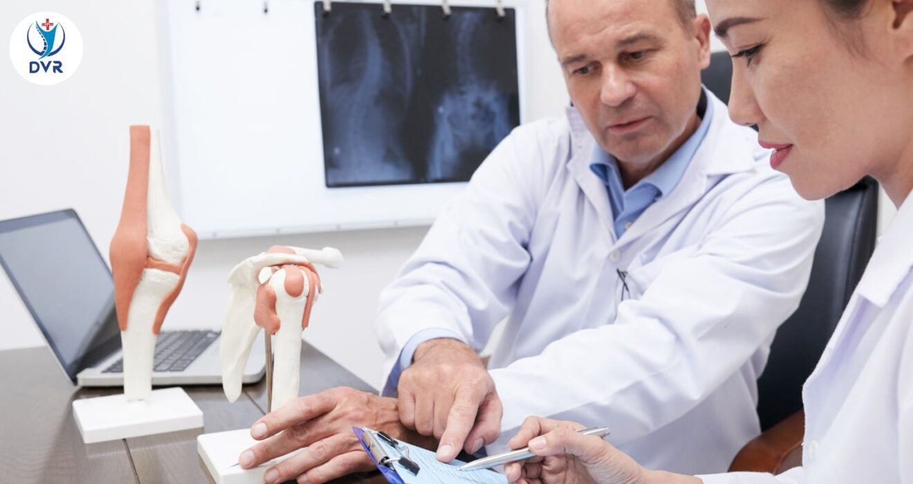
Joint pain can significantly affect daily life, limiting movement and causing discomfort during routine activities. Whether it’s persistent knee pain, shoulder stiffness, or unexplained swelling in the wrist or ankle, accurate diagnosis is the first step toward effective treatment. Among modern imaging technologies, MRI (Magnetic Resonance Imaging) has become one of the most reliable methods for identifying joint problems.
Many patients today prefer visiting a diagnostic centre in narsingi for advanced imaging because of the availability of modern MRI machines and expert radiology support. MRI scans provide highly detailed images of bones, cartilage, ligaments, tendons, and soft tissues, helping doctors detect issues that other imaging techniques may miss.
This guide explains how MRI scans work for joint issues, what conditions they detect, and why choosing a reliable diagnostic centre in narsingi can help in getting accurate results for proper treatment planning.
Understanding MRI Scans for Joint Evaluation
An MRI scan uses strong magnetic fields and radio waves to create detailed images of the body’s internal structures. Unlike X-rays or CT scans, MRI does not use radiation. This makes it a safe and effective diagnostic tool for examining joints and surrounding tissues.
Doctors often recommend an MRI when a patient experiences:
- Persistent joint pain
- Swelling or inflammation
- Limited range of motion
- Suspected ligament or tendon injury
- Unexplained joint instability
At a professional diagnostic centre in narsingi, MRI scans are performed using advanced imaging systems that capture highly precise images. These images help orthopedic specialists understand the exact cause of joint problems and decide the most suitable treatment approach.
Why MRI is Important for Joint Problems
Joint issues can originate from multiple structures including bones, cartilage, ligaments, tendons, and surrounding muscles. Traditional imaging methods sometimes fail to detect subtle damage in these soft tissues.
MRI scans provide several advantages:
Detailed Soft Tissue Imaging
MRI produces high-resolution images that clearly show soft tissues. This helps doctors identify ligament tears, cartilage damage, and tendon injuries that might not appear on X-rays.
Early Detection of Problems
Certain joint conditions, such as cartilage degeneration or early arthritis, can be detected at an early stage through MRI scans performed at a trusted diagnostic centre in narsingi.
Non-Invasive Procedure
MRI scans do not require surgery or invasive procedures. Patients simply lie inside the scanner while images are captured.
Accurate Treatment Planning
By identifying the precise cause of joint pain, MRI helps doctors plan treatments such as physiotherapy, medication, or surgery.
Common Joint Conditions Detected by MRI
MRI scans can detect a wide range of joint problems. Below are some of the most common conditions that doctors diagnose through MRI imaging.
Ligament Injuries
Ligaments connect bones and help stabilize joints. Injuries to these structures are common in sports or accidents.
MRI can detect:
- ACL and PCL tears in the knee
- Sprains in the ankle
- Ligament injuries in the wrist or elbow
Visiting an advanced diagnostic centre in narsingi ensures that such injuries are detected with high accuracy.
Cartilage Damage
Cartilage is a flexible tissue that cushions joints and prevents bones from rubbing against each other. Damage to cartilage can cause pain, stiffness, and limited mobility.
MRI scans can identify:
- Cartilage thinning
- Cartilage tears
- Early degeneration
Early detection at a reliable diagnostic centre in narsingi allows doctors to recommend treatments that prevent further joint deterioration.
Tendon Injuries
Tendons connect muscles to bones and help joints move smoothly. Overuse, injury, or aging can lead to tendon problems.
MRI scans help detect:
- Tendon inflammation (tendinitis)
- Partial or complete tendon tears
- Degenerative tendon changes
Accurate imaging from a professional diagnostic centre in narsingi helps orthopedic specialists determine whether surgery or conservative treatment is required.
Meniscus Tears
The meniscus is a cartilage structure in the knee that acts as a shock absorber. Meniscus tears are common in athletes and older adults.
MRI scans can clearly show:
- Meniscus tears
- Degenerative meniscus damage
- Associated ligament injuries
Since knee injuries are complex, doctors often rely on imaging results from a trusted diagnostic centre in narsingi for accurate diagnosis.
Arthritis and Joint Degeneration
Arthritis is one of the leading causes of joint pain worldwide. MRI scans help detect different types of arthritis at early stages.
MRI can identify:
- Rheumatoid arthritis
- Osteoarthritis
- Joint inflammation
- Bone erosion
Early diagnosis through imaging at a reliable diagnostic centre in narsingi helps patients start treatment sooner, reducing long-term joint damage.
Bone Abnormalities
Although MRI is primarily used for soft tissue imaging, it can also detect problems within the bone.
MRI scans can reveal:
- Bone fractures not visible on X-rays
- Bone infections
- Bone tumors or cysts
- Bone marrow abnormalities
Because of its high sensitivity, MRI is often recommended when other tests cannot determine the cause of joint pain.
Joints Commonly Examined with MRI
MRI scans are widely used to examine several joints in the body. Some of the most frequently scanned joints include:
Knee Joint
The knee is one of the most commonly injured joints. MRI scans help diagnose ligament tears, cartilage damage, meniscus injuries, and arthritis.
Shoulder Joint
Shoulder pain may result from rotator cuff injuries, tendon inflammation, or joint instability. MRI provides clear images of these structures.
Hip Joint
MRI helps detect hip joint issues such as cartilage damage, labral tears, and early arthritis.
Wrist and Hand
Small joints in the wrist and hand require highly detailed imaging, which MRI provides effectively.
Ankle and Foot
MRI scans can detect ligament injuries, tendon tears, and bone abnormalities in the ankle and foot.
Patients visiting a well-equipped diagnostic centre in narsingi can undergo MRI scans for any of these joints with accurate results and minimal waiting time.
What to Expect During an MRI Scan
Many patients feel anxious about MRI scans because they are unfamiliar with the procedure. However, the process is simple and painless.
Preparation
Before the scan, patients may be asked to remove:
- Metal objects
- Jewelry
- Watches
- Electronic devices
The technician will explain the procedure and position the patient comfortably on the MRI table.
During the Scan
The table slides into the MRI machine, where magnetic fields create detailed images. The scan usually takes 20 to 45 minutes depending on the joint being examined.
Patients are required to remain still during the scan to ensure clear images. Some machines may produce loud sounds, but ear protection is typically provided.
At a professional diagnostic centre in narsingi, trained technicians ensure that the scanning process is smooth and comfortable.
After the Scan
Once the scan is completed, patients can resume normal activities immediately. The images are reviewed by a radiologist who prepares a detailed report for the doctor.
Who Should Consider an MRI for Joint Pain
Doctors usually recommend MRI scans when:
- Pain persists for several weeks
- Physical therapy does not improve symptoms
- X-rays do not reveal the problem
- There is suspected ligament or tendon injury
- Surgery is being considered
Getting an MRI done at a trusted diagnostic centre in narsingi ensures that doctors receive accurate imaging information before deciding on treatment.
Benefits of Choosing a Reliable Diagnostic Centre
Selecting the right diagnostic facility plays a crucial role in obtaining precise MRI results. High-quality imaging equipment and experienced radiologists contribute to accurate diagnosis.
A good diagnostic centre in narsingi typically offers:
- Advanced MRI technology
- Experienced radiology specialists
- Quick appointment scheduling
- Detailed and reliable reports
- Comfortable scanning environment
Accurate imaging results help doctors create effective treatment plans, reducing the risk of misdiagnosis.
Safety of MRI Scans
MRI scans are generally considered very safe because they do not use ionizing radiation. However, patients should inform the technician if they have:
- Pacemakers
- Metal implants
- Cochlear implants
- Certain surgical clips
The medical team at a professional diagnostic centre in narsingi will review the patient’s medical history to ensure the procedure is safe.
When Early Diagnosis Makes a Difference
Joint problems often worsen when ignored. Conditions like ligament tears, cartilage damage, and arthritis can progress if not treated early. MRI scans allow doctors to identify these issues at an early stage.
Early detection offers several benefits:
- Faster recovery
- Less invasive treatment
- Reduced long-term joint damage
- Better mobility and quality of life
This is why many healthcare professionals recommend timely imaging at a reliable diagnostic centre in narsingi when joint pain persists.
How MRI Helps Diagnose Different Types of Joint Pain
Joint pain is one of the most common health concerns affecting people of all age groups. It can occur due to injuries, aging, inflammation, or underlying medical conditions. When pain becomes persistent or affects mobility, doctors often recommend advanced imaging tests to determine the exact cause. One of the most effective diagnostic tools for evaluating joint conditions is the MRI scan.
An MRI scan provides detailed images of bones, cartilage, ligaments, tendons, and surrounding soft tissues. This makes it extremely useful in diagnosing complex joint problems that may not be visible through other imaging methods. Many patients today prefer visiting a diagnostic centre in narsingi for MRI scans because these facilities are equipped with advanced imaging technology and experienced radiology teams.
Understanding how MRI works and how it helps diagnose various joint problems can help patients make informed healthcare decisions.
Understanding Joint Pain and Its Causes
Joint pain can arise from multiple structures within the joint. Since joints consist of bones, cartilage, ligaments, tendons, and fluid-filled sacs, identifying the exact source of pain can sometimes be challenging.
Common causes of joint pain include:
- Sports injuries
- Ligament sprains or tears
- Cartilage damage
- Arthritis and inflammation
- Tendon injuries
- Bone fractures
- Joint infections
In many cases, doctors initially recommend physical examinations or basic imaging like X-rays. However, if the problem is related to soft tissues or early-stage degeneration, an MRI scan performed at a trusted diagnostic centre in narsingi can provide more detailed information.
Why MRI is Preferred for Joint Diagnosis
MRI technology is widely used in orthopedics because of its ability to capture high-resolution images of both bone and soft tissue structures.
Detailed Imaging of Soft Tissues
Unlike X-rays, MRI scans provide clear images of soft tissues such as ligaments, tendons, and cartilage. This helps doctors detect injuries that are otherwise difficult to identify.
Patients who undergo imaging at a reliable diagnostic centre in narsingi often benefit from accurate diagnosis due to advanced MRI systems.
Early Detection of Degenerative Conditions
Many joint diseases develop slowly over time. MRI scans can detect early signs of cartilage wear, bone changes, or inflammation before the condition becomes severe.
Non-Invasive and Safe
MRI scans do not use radiation, making them a safe diagnostic option for many patients. The procedure is painless and usually completed within a short time.
Types of Joint Problems MRI Can Diagnose
MRI scans can help doctors detect several types of joint conditions. Below are some of the most common problems identified through MRI imaging.
Ligament Tears and Sprains
Ligaments are strong bands of tissue that connect bones and stabilize joints. Injuries to ligaments are common in sports and accidents.
MRI scans can detect:
- Partial ligament tears
- Complete ligament ruptures
- Ligament stretching or sprains
For example, ACL injuries in the knee are often diagnosed using MRI scans performed at a well-equipped diagnostic centre in narsingi.
Early detection of ligament injuries helps doctors recommend appropriate treatments such as physiotherapy, bracing, or surgery.
Cartilage Injuries
Cartilage acts as a cushion between bones in a joint. Damage to cartilage can cause pain, stiffness, and reduced mobility.
MRI scans help identify:
- Cartilage thinning
- Cartilage cracks or tears
- Early cartilage degeneration
Patients experiencing chronic knee or hip pain often undergo MRI scans at a diagnostic centre in narsingi to evaluate cartilage health.
Early diagnosis can prevent long-term joint damage and help doctors recommend protective therapies.
Tendon Disorders
Tendons connect muscles to bones and play a vital role in joint movement. Tendon injuries may occur due to overuse, sports activities, or age-related degeneration.
MRI scans can reveal:
- Tendon inflammation
- Tendon tears
- Tendon degeneration
For example, shoulder pain caused by rotator cuff injuries is frequently diagnosed using MRI imaging available at an advanced diagnostic centre in narsingi.
Meniscus Tears in the Knee
The knee joint contains two cartilage structures called menisci that act as shock absorbers. Meniscus tears are among the most common knee injuries.
MRI scans provide detailed images that help doctors identify:
- Horizontal meniscus tears
- Vertical meniscus tears
- Degenerative meniscus damage
Accurate diagnosis at a trusted diagnostic centre in narsingi helps orthopedic specialists decide whether the patient needs physiotherapy, medication, or surgery.
Arthritis and Inflammatory Conditions
Arthritis is a major cause of chronic joint pain. MRI scans are particularly helpful in identifying different forms of arthritis at an early stage.
MRI imaging can detect:
- Joint inflammation
- Synovial membrane swelling
- Cartilage loss
- Bone erosion
Patients who experience persistent stiffness or swelling may benefit from early MRI evaluation at a professional diagnostic centre in narsingi.
Early treatment can slow disease progression and improve joint function.
Bone Injuries and Hidden Fractures
While X-rays are commonly used to detect fractures, some bone injuries are difficult to identify through standard imaging.
MRI scans can detect:
- Stress fractures
- Bone bruises
- Bone marrow abnormalities
- Micro fractures
These conditions may be diagnosed accurately through imaging performed at a reliable diagnostic centre in narsingi.
Joints Frequently Evaluated with MRI
MRI scans are commonly used to evaluate several major joints in the body.
Knee
The knee is one of the most complex joints and is highly prone to injuries. MRI helps diagnose ligament tears, cartilage damage, and arthritis.
Shoulder
Shoulder MRI scans help detect rotator cuff injuries, tendon inflammation, and joint instability.
Hip
Hip MRI scans are useful for detecting cartilage damage, labral tears, and early arthritis.
Ankle
MRI scans can reveal ligament injuries, tendon damage, and bone abnormalities in the ankle joint.
Wrist
Wrist MRI scans help diagnose ligament injuries, small fractures, and tendon disorders.
Patients visiting a diagnostic centre in narsingi can undergo MRI scans for any of these joints depending on their symptoms.
When Doctors Recommend an MRI Scan
Doctors may suggest an MRI scan if a patient experiences:
- Persistent joint pain
- Swelling that does not improve
- Reduced joint mobility
- Joint instability
- Suspected ligament or tendon injury
In such cases, getting the scan done at a trusted diagnostic centre in narsingi ensures high-quality imaging results for accurate diagnosis.
What Happens During an MRI Scan
Many people feel nervous before undergoing an MRI scan, but the process is simple and comfortable.
Before the Scan
Patients may be asked to remove metal items such as jewelry, watches, or belts. The technician will explain the scanning procedure and answer any questions.
During the Scan
The patient lies on a scanning table that moves into the MRI machine. The scanner uses magnetic fields and radio waves to create images of the joint.
The procedure usually takes about 20 to 45 minutes.
Technicians at a professional diagnostic centre in narsingi ensure that patients remain comfortable during the entire process.
After the Scan
Once the scan is completed, patients can return to their normal routine. The images are reviewed by a radiologist who prepares a detailed report for the referring doctor.
Benefits of Early MRI Diagnosis
Early diagnosis plays a crucial role in treating joint problems effectively.
Some of the major benefits include:
- Accurate identification of the problem
- Faster recovery with proper treatment
- Prevention of long-term joint damage
- Reduced risk of surgery in some cases
By choosing a reputable diagnostic centre in narsingi, patients can ensure that they receive precise imaging results that support effective medical care.
Choosing the Right Diagnostic Centre
The quality of MRI results depends heavily on the technology used and the expertise of the radiology team.
A reliable diagnostic centre in narsingi usually offers:
- Advanced MRI machines
- Experienced radiologists
- Accurate diagnostic reports
- Comfortable patient facilities
- Quick appointment scheduling
These factors contribute to better diagnostic accuracy and improved patient outcomes.
Safety Considerations for MRI Scans
MRI scans are generally safe for most patients because they do not use radiation. However, patients should inform medical staff if they have:
- Metal implants
- Pacemakers
- Artificial joints
- Cochlear implants
Medical professionals at a trusted diagnostic centre in narsingi carefully evaluate each patient’s medical history before performing the scan.
Conclusion
Joint pain can result from many different conditions, ranging from minor injuries to chronic diseases. Identifying the exact cause of the problem is essential for proper treatment. MRI scans have become one of the most reliable diagnostic tools for evaluating joint health because they provide detailed images of bones, cartilage, ligaments, and soft tissues.
By visiting a trusted diagnostic centre in narsingi, patients can benefit from advanced MRI technology and expert radiological analysis. Accurate imaging not only helps doctors diagnose joint problems more effectively but also allows them to design the most appropriate treatment plan for faster recovery and improved mobility.
Contact Us:
Call us on : 093910 29909
Follow Us On Instagram : https://www.instagram.com/dvrhospital/
Follow Us On Facebook : https://www.facebook.com/people/DVR-Hospital-Diagnostics/61580485479021